

 Passport






Mastodons of the UW Geology Museum
01/01/70 | 35m 56s | Rating: NR
Carrie Eaton, Curator of the Geology Museum at UW-Madison, shares the story behind a black and white photograph showing a group of men holding the femur of a massive ice-age behemoth. A visitor brought the photo to the UW’s Geology Museum hoping to learn the whereabouts of the bone. Learn how Eaton solved the mystery of where the photo was taken.


Copy and Paste the Following Code to Embed this Video:
Mastodons of the UW Geology Museum
Welcome everyone to Wednesday Night at the Lab, except this is a special Friday night edition of Wednesday Night at the Lab. We're here to launch the 14th annual UW-Madison Science Expeditions campus-wide open house. So thank you very much for coming to Science Expeditions. It's the open house for Your Public Land-Grant Research University.
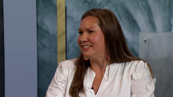
Tonight I'm delighted to introduce to you Carrie Eaton. She's with the UW-Madison Geology Museum. She'll be here to talk about the mastodons at the UW Geology Museum. Carrie was born in Woodridge, Illinois, and graduated from Downers Grove South High School in Illinois.
Then she went to the University of Iowa to study geology and then she came here to get her master's degree in Quaternary Geology, Quaternary. -
Voiceover
Qua-ter-na-ry. - Qua-ter-na-ry. See, the chemists pronounce it different than the geologists. -
Voiceover
It's true. I always loved the word, I never could get it out straight. She's been with the UW Madison Geology Museum since 2009, but the Geology Museum has been around about 1848. It is older than the university, and there aren't very many things at the university that are older than the university.
And it's one of the splendid places. They're gonna have their open house as part of Science Expeditions. They're open six days a week nearly every week of the year. And if you get a chance to come and visit the Geology Museum, I think you'd be very pleasantly surprised.
So please join me in welcoming Carrie Eaton to Wednesday Night at the Lab here for UW Science Expeditions. (audience applauds) Thank you. Thank you, Tom, and thank you, everyone, for being here tonight. Here we go.
Yeah, so I'm the curator of collections at the UW Geology Museum, and I'm going to be talking about our mastodons tonight and how they're related to the university history, but also how they're linked to the history of our state, and in a way to the history of science itself. As Tom mentioned during the introduction, the UW Geology Museum is itself also kind of an old place. We got our start in 1848 at the very first meeting of the Board of Regents. They decided they wanted to form a mineral cabinet, cabinet being the Victorian word for museum or collection.
And once they had acquired enough specimens, they had enough to open up a public museum. For 65 years, this public museum had its home in Science Hall, and as the collection grew it included things like birds and mammals that would become part of a zoology collection, and eventually also some plants and other herbarium specimens that would later become the Wisconsin State Herbarium. Eventually the Geology Museum was able to put on some large skulls and skeletons on display, including this one here, which is the skeleton of another ice age mammal called a Giant Ground sloth. Eventually this ground sloth was taken down and in its place they put up this specimen of a mastodon.
This mastodon was installed in 1915. So in the fall of last year it celebrated 100 years on display. Now, mastodons are just one of 37 different genera of large mammals that went extinct at the end of the Pleistocene epoch, the geological period of time that we all commonly think of as the Ice Age. Now, in addition to some of these large mammals that went extinct, there were some other large animals that were locally extirpated from the regions in which they live, and now, by and large, only exist in places that are generally pretty cold and dry.
Mastodons roamed North America from about 3.75 million years ago until their extinction at the end of the Ice Age about 11,700 years ago. They were spruce forest dwellers, so they lived in places that looked like this, and they used their cusped teeth to crush things like needles, leaves, twigs, woody and brushy plants. Now, the very first skeleton of a mastodon was excavated in 1801 and it was painted by the person behind the endeavor, Charles Willson Peale. This painting called the Exhumation of the Mastodon was painted in 1806.
And pictured over here on the right-hand side, you can actually see Charles Willson Peale and his son, Rembrandt, who was also an artist, over here with the family while the laborers pump the water out of the hole to be able to excavate the mastodon skeleton. Now, when this mastodon skeleton was exhumed, it became the first fully-articulated skeleton of a mastodon to go on display, and was actually the second skeleton to be on public display in a museum anywhere. Now if we look at all of the mastodon localities in the upper Midwest, you can see that mastodons are pretty prevalent here as well. And if we look at Wisconsin in general, you'll see that all of those little blue dots that indicate mastodon sites are scattered across the southern half of the state.
Now, this makes sense because throughout the Pleistocene or the Ice Age there were several glacial readvances and retreats that occurred in the northern portion of the state. Mastodons by and large relied on the presence of spruce forest, so as the glaciers retreated and as the spruce forest migrated into the southern part of the state, they had all of the kinds of flora and fauna that they needed for their regular habitat. Now, one of these mastodon finds is pretty well-known. In the summer of 1897, a group of young boys, four brothers on a farm, discovered a mastodon in Richland County.
Every school group that has come to our museum for the past several decades has heard the story of this mastodon. They've sat at its feet and they heard how after heavy rains these four brothers went out on their farm to inspect a floodgate, and they found some large bones protruding out of a stream bank. So for many decades, this specimen has been known to all, and to some who still come to our museum today, as the Boaz mastodon. But when I started digging into the mastodon's history in preparation for the 100th anniversary, I discovered something kind of unusual.
And so in collaboration with David Null who is the director of UW Archives, we started going through some old university correspondence and university records, and we came upon this particular letter that I found very curious. And it describes the purchase of a mastodon that was found near Fennimore in Grant County, Wisconsin. And this letter I particularly liked because it even described that assistant state geologist, "Mr. Ernest Buckley purchased this skeleton "for the university knowing that Professor Van Hise," that's Charles Van Hise, who would later become president of the UW, "was desirous of obtaining enough bones in order "to reconstruct a complete skeleton." Now, this letter was written by E.
A. Birge and it was sent to the secretary of the Board of Regents, and it was written in the summer of 1898, the very summer after the Boaz mastodon had been excavated and purchased by the university. So this was kind of interesting. Now, this mastodon was pretty well-represented in historical newspaper accounts, as this mastodon that was discovered near the small hamlet of Anderson Mills.
It was found by a farmer's young son. Harry Anderson was going out to the field getting ready to work one morning after heavy rains and found something protruding out of the ground. Further digging revealed several bones of a specimen of a mastodon. Now, a number of years ago, a visitor to the museum happened to bring us some photographs of this very mastodon find, wondering if we knew anything about it.
So this is actually a picture of the Andersons out in front of their farm home, with the bones of their mastodon sitting at their feet. One of the other photographs portrays assistant state geologist Ernest Buckley, along with some of the local farmhands and rail workers who he convinced to help him with the excavation. And yet another photograph shows these bones piled up at the foot of a tree somewhere on the family's property. So we decided we really needed to take a closer look at these photographs.
And one in particular drew our attention, and it was this picture of assistant state geologist Ernest Buckley holding the femur of the mastodon. And what we noticed, the femur is actually upside down in this picture, so the head of the femur is down toward his feet, and we're actually looking at the back of the femur, or thigh bone, so that the back of the knee is up near his shoulder. Now, where Mr. Buckley's left hand is sitting on the bone there should be something called a condyle, a lateral condyle or a condyle towards the outside of the body.
And that condyle is missing. Now, just above his right hand below his right shoulder, there is a rounded portion of bone called a condyle, and that condyle is present. And so we wanted to take a closer look at this particular bone, and see if we noticed it in any of the other photographs. And sure enough, because if you're gonna get your picture taken with some mastodon bones, you should grab the biggest one you can find.
There's a picture of Harry Anderson standing holding the femur of the mastodon. And this is a left femur. And so up here you can see the head of the femur, what would articulate with the hip socket, and this time we're actually looking at the front surface of the bone, or the anterior surface. And down here where you would see what's called the patellar groove, and that's what helps to make your knee a hinge joint.
This would articulate with what becomes your knee. Down here, the patellar groove is completely broken away, and you can see that there's a fracture running through the front surface of this bone. So one really great way to inspect bones more closely is to collaborate with the Wisconsin Institutes of Medical Research and convince them to let you use their medical CT scanner. So we actually got to put our mastodon femur through a very high resolution CT scanner over at the UW CT lab.
And the reason that we did this is because it really helped us to see past all of the plaster reconstruction, and paint, and varnish that had been put on this bone in order to prepare it for display more than 100 years ago. So what we're actually looking at is a video of CT slices. So if you could imagine looking through a loaf of bread, slice by slice, and the scan is actually oriented so that the head of the femur, what would articulate with the hip, is up at the top of our video, and the back of the knee is down at the bottom. I'll see if we can play this one again so you can see it really well.
Now, what helps to illustrate this even more is if we apply a color gradient to it. And so real bone is appearing as a kind of purple-blue color, and this bright green that we're seeing is the plaster reconstruction. You'll notice that that bright green is also filled with little black holes, and those little black holes are air pockets that form as the plaster dries. We'll see if I can go back and play it again.
Here we go. So you'll see again our purple bone up in the head of the femur, and again in that medial condyle where we could see in that photograph that that was present, the lateral condyle is made entirely of plaster, as is the anterior portion of the distal end of the femur or what would become the front of the knee. So basically the CT scan showed us exactly what we were seeing in the photographs, that this femur was a match to the photographs of what we were seeing in Anderson Mills. Now, the historical newspaper accounts also describe the specimen coming to the university and being studied by people in Science Hall.
And one of the things that they noticed once the bones of the skeleton came here, was that it seemed as though the animal had once had two broken ribs. And I love the way they describe this, "The knitting of the parts being very apparent." And that's because it perfectly describes exactly what you would see. And so sure enough, we went out to look at our skeleton on display in the museum. And you can see up here, in what are the 18th and 19th ribs on the animal's left-hand side, there are these two bulbous regions at approximately the mid-shaft portion of the rib, and if we take them off display you can see it even better.
And sure enough, you can see these kind of large, bulbous portions on these bones that are very consistent with a bone having been broken and healed during the animal's lifetime. So we had our two broken ribs as well, indicating again that we had more skeletal elements that hailed from the Anderson Mills site. So if we go back to our bone pile photograph, we could look through one by one and pick out elements that were present in the photograph that matched elements that were in our skeleton on display. So this really started to paint a pretty consistent picture of many of the bones belonging to the Anderson Mills site.
What's more, when we identified which elements were coming from Anderson Mills, we noticed some similarities in their coloration and also the presence of this particular staining. So you can see here that there is a slightly dendritic or veined pattern on the back side of these bones. We're looking at the back side of the left tibia here on the left-hand portion of the screen, and we're looking at the back of the ribs on the right-hand portion of the screen. You'll notice that the bones all have this brownish honey color and they have this dendritic staining.
This staining is indicative of a low oxygen or anoxic environment. And what's more, this staining only seemed to occur on one side of the bone. So again, very consistent. These bones all seemed to be hailing from the same site.
They had very similar coloration, they had very similar quality of preservation, and they had similar taphonomy. And taphonomy just describes what happens to a bone after it's been deposited. So this had me wondering, "Well, if most of this "is coming from Anderson Mills, "then what has happened to the Boaz mastodon?" And so I went to the Wisconsin Geological and Natural History Survey and I poured through the historical field notebooks that they have kept on file. And thank goodness, because I was able to find one of Ernest Buckley's field notebooks.
So in a field notebook labeled Observations to Be Made On Quarries, Cautions to Be Observed, Materials to Be Collected, Et Cetera, I found in the summer of 1897 a map showing the position of bones found, and this looked strikingly like a portion of a stream with some large bones drawn into its bank, and a small little stratigraphic section showing the kind of soil that was found at the site. Also included was a list of bones that had been found. Now, Buckley's list was by and large describing things that sounded small or fragmentary. There were fewer than 30 pieces at the site.
Most of them were described as having been broken bones or pieces of bones. Only one rib was described as being nearly whole, and only one vertebra was described as being nearly complete, when we had more than 20 intact vertebrae in our skeleton and more than 34 complete ribs. So this list could not possibly account for all of the genuine bones that were present in the skeleton that we had on display. So after we grouped all of the bones together by what we could see in the photographs and what we were able to tell by the characteristics that they shared, that coloration, that staining, that taphonomy, we were left with two outliers that we could attribute to the Boaz site.
So what we're looking at here, a picture here on the left is a right tibia, and on the right-hand side is the left first rib. And so one way to tell whether or not these skeletal elements were coming from two different animals was to drill into them and collect core subsamples that we could then use for radiocarbon analysis. This would give us the general idea of a date of the specimen, and it would tell us whether or not these bones seem to be coming from a similar time or from different time periods. So when we got our radiocarbon results back they clustered into two very different groups.
And so what we're looking at here are radiocarbon dates and they are represented here on a horizontal axis that shows calendar years before present. And each of these elements is labeled based on where it came from. And so what you'll notice here is that our left first rib and right tibia are clustering together, and the left femur and the left humerus are clustering together. Now, the dates that we're looking at here, these horizontal bars here that are colored in solid red, this shows the radiocarbon date with a 68% confidence interval.
Now, if we want to be extra confident, we go all the way out to the end of these whiskers and this shows us the date range with a 95% confidence interval. Now, this curvy line that's been shaded in above our horizontal bars, this is something called the 100% probability curve, and it shows us the probability that the radiocarbon date is occurring within this range. The most important thing that you should notice about this plot is that these curves do not cross and these dates do not overlap. In fact, if we look at the commonality of where these dates do overlap by their group, we see that these two sets of bones are separated by about 750 years.
So the younger of the two, the two that we attribute to the Boaz site, our left first rib and our right tibia, they come in at about 12,150 calendar years before present. Now, the others, the left femur and left humerus attributed to the Anderson Mills site have a radiocarbon date of about 12,900 calendar years before present. So if we then take that information and look at the skeleton as a whole, we can see that by and large the majority of the skeleton likely comes from the Anderson Mills site. With more than 60 complete elements it makes up the majority of the genuine bones that we have on display.
But what's interesting about that radiocarbon date from the Boaz site is that it is actually one of the youngest radiocarbon dates on a mastodon in the upper Midwest, which means that the Boaz mastodon could have been one of the latest mastodons to be in this region, which places it among some of the last mastodons standing before the large extinction event at the end of the Pleistocene epoch. So that's pretty neat. Now, because we had so much of the Anderson Mills skeleton present, that means that we could actually look at the animal and tell a little bit more about its life history. So the kinds of things I wanted to figure out was whether or not we could tell the specimen's gender and whether or not we could figure out how old it was.
And so one really great way to do that is actually to look at the animal's teeth. So males, by and large in mammals, tend to be generally larger than females. So one way you can compare size is by looking at the last molar that's present in the mastodon's mouth, or what's called the M3, and by measuring its size. And we do that by measuring its maximum length versus its maximum width.
So taking those measurements and comparing that to other known specimens that are out there, it seemed as though the Anderson Mills mastodon was probably male. Now, the way we know that tooth is the last molar is by counting the number or ridge crests or what are called loafs. So mastodon teeth have these rows of what look like cusps, is what we call them ridge crests. And those ridge crests are called loafs.
And the very last molar that a mastodon will ever get in its mouth will have five of these ridge crests or loafs on it, which means that the Anderson Mills mastodon was also a mature adult. Now, based on some of the other things that we've seen in the skeleton, there are small bone lesions and tubercles that appear on some of the bones. There are also some rather gnarled attachment points where tendons and muscles would attach. It seems as though the mastodon was probably even a little bit older, so that would place it at at least 30 years old, and based on some of the other things we've seen, probably a little older than that.
Another great way to determine gender is by looking at the mastodon's femur. And so with femur or thigh bones, there tends to be a pretty big discrepancy between males and females. You can tell them apart pretty easily by looking at size. And so if we take a more generous measurement of the femur and we measure its entire articular length, it comes in at 107.5 centimeters.
That's about three feet long. We can also use a more conservative measurement and measure it only from the edge of that lateral condyle up to what's called the greater trochanter, that ridge that I've outlined there, and that length is about 98 centimeters. That's still pretty long. You can then also measure the midshaft circumference on the femur, and this one comes out at about 39 1/2.
So mastodon females have femoral lengths, or length of their femur that tends to be in the 90's. So when we compare this one to the specimens that are published in the known literature, it is most definitely a male. If most females are clocking in somewhere between 85 to 95 centimeters for their total length, this one actually turns out to be a pretty big mastodon, which pretty well ensures that it is, in fact, male. Now, the reason we had to use these measurements, looking at the teeth and also looking at the femur of the mastodon, is because we only had a partial pelvis.
So the pelvis is always the easiest place to start, but if that's not complete, you can use these other methods in order to determine gender and age. But the story doesn't end there because the very day after the Anderson Mills mastodon was discovered, a conductor on the narrow-gauge rail line that ran through that region put a few of the elements on display at the Fennimore Depot in order to share the news with the local citizens. And sure enough, this is described pretty well in the historical newspaper accounts, that Conductor Gifford exhibited two specimens of the skeleton, "A rib 3 1/2 feet long and a vertebra." And so I was quite surprised and rather delighted when last summer I got a phone call from the mayor of Fennimore, who also happens to be an officer of the Fennimore Historical Society's Railroad Museum that's in that town, telling me that they had bones of a mastodon. And I said, "Oh, really?" (laughing) And so those bones actually came to visit our museum.
And I was pretty nervous and quite excited. And when he brought them in and we got to unwrap them I was also quite relieved because they were, in fact, bones from the Anderson Mills mastodon. And so sure enough, we have a vertebra measuring roughly 10 inches across, and a rib roughly 3 1/2 feet long. The quality of preservation was identical to the bones that we had in our mastodon.
The coloration and even the presence of the staining on one surface of the bone. What's more, is that when we look at these bones, what's a first lumbar vertebra and a rib from the right-hand side, we looked where those bones should be on our skeleton, we had replicas. So seems like these are most definitely some bones of the Anderson Mills mastodon. They were actually kept by Conductor Gifford and then they were passed on through his family.
They were eventually handed over to the Roethe family who then became owners of the Fennimore Times Review. They were donated to the Railroad Museum in 1993. So when they came for their visit we did our due dilligence in photographing them and measuring them as thoroughly as we could, and we returned them to the museum. So they will be on display at the Railroad Museum and the folks in Grant County will be able to go to the Railroad Museum and see bones of their local mastodon on display.
But the historical newspaper accounts describe something else, and that's that Harry Anderson found a tusk. The very first element recovered from this mastodon was that of a tusk, "Measuring nine feet four inches "in length with a circumference at the base of 25 inches." That's a pretty big tusk which would be consistent with it being a male. And people always ask me, "What happened to the tusk? "What happened to the tusk?" Well, tusks will exfoliate very rapidly once they've been underground and they're brought to the surface.
If they're not embedded with adhesives or preserved right away, they have this tendency to crack as they dry and fracture apart. So just a few months ago, I got a really neat phone call from someone who happens to be an Anderson relative. That phone call was followed up by a pretty cool email that had an attachment to it. (laughing) And so, I notice right away that what we had here were definitely some pieces of what could be teeth or enamel.
And what's more, included in the picture was what looked like an original copy of the Fennimore Times Review newspaper article. So when the box arrived at my office, it was clear that we were dealing with something that had been in someone's family for a very long time. And when I opened the box, there were, in fact, some pieces of enamel. These brown fragments that you're seeing here are actually the exterior surface of a tusk.
And these paler, whiter fragments are portions from the interior of the tusks. So what you're seeing is the difference between a polished and mineralized enamel, and the dentin that makes up the interior of the tusk, because tusks, of course, are just modified teeth. And folded neatly inside was the original copy of the Fennimore Times Review newspaper article, so if we zoom up closely, you can see it says, "Unearthed mastadon bones, "skeleton now in possession of the state." So that's pretty neat, someone from the family neatly cut along the column to preserve this article. And a second layer of tissue paper revealed an even larger fragment from the interior portion of the tusk, measuring somewhere in between about five to six inches in length.
All of these fragments and the newspaper article are now part of the UW Geology Museum's permanent collection. So it was pretty neat for us to welcome these pieces back home, and reunite them with the rest of the skeleton. Now studying this mastodon also gave us a unique opportunity to attempt something new with preservation, and conservation, and replication of bones. So our museum scientist Dave Lovelace spearheaded a rather rigorous effort to digitally scan the surface of our mastodon's femur.
And you can see on the surface all of these little white reference marks. They allowed him to piece together all of those individual digital scans, and made sure that the surface was referenced and matching the same volume of the bone that we had on display. Now, you can do this also by surface rendering from a CT scan, which is what I've done here. So I've taken the CT scan data and I've created a surface model of it.
But you'll notice here that the surface model is based on density, and so we're seeing holes where there is some of that spongy bone present. We're also seeing some holes where there are fractures or cracks in the surface, or maybe there's been plaster reconstruction, so it's not appearing as dense. Using this would require quite a bit of smoothing. And now we actually have some software that helps us use the CT data to make surface models.
But so this is why we went with the 3D digital scanner instead. And so with some very rigorous scanning of the surface, Dave and one of our museum students, James, were able to create a new 3D surface model and actually begin printing the mastodon femur on our 3D printer. And so piece by piece we would print off sections of slices and then put them together to create a whole. Then what was really cool is we realized that we had an opportunity here to create a copy of our mastodon's femur without ever touching the bone itself.
Now, I was tasked with having to drill into a bone that had been on display for 99 years to collect subsamples for radiocarbon dates. That was nerve-wracking enough. And as you've seen from the pictures of the tusks, bones that have been in the ground for a long time and have been out of the ground for a long time have a tendency to fracture and break. So a bone that is both old and has been on display for a long time often doesn't handle rigorous treatment very well.
So what we actually did is Dave created a mold, a mother mold, of the 3D printed femur. And from that mold we were able to create another replica. So we're reproducing mastodon femurs without ever actually laying hands on the bones using only technology that is available to us at the museum and here at the Wisconsin Institutes for Medical Research. And so I actually brought the femur with me today.
(laughing) And so this is what it looks like when you 3D print a mastodon femur. And it also gives you a sense of perspective for how large this animal was. As you can see, it's thigh bone is taller than my waist. And what was really great is that even though we can see that this was 3D printed, we actually got some really great fidelity, and you can see a lot of the structures present in the original bone that are in fact present on our 3D replica here, including this very characteristic fracture on the anterior surface of the bone showing the difference between the genuine bone and that plaster reconstruction.
And in fact, you can almost make out the difference between the genuine medial condyle and the reproduced lateral condyle. So the fidelity was good enough that we were even picking up some of those clues that led us to our discovery in the first place. And I will leave this here and I would welcome all of you to check it out at the end of my talk. He kinda goes with me everywhere.
(laughing) And so what this brings me to is that some of you in the audience might be wondering, "What's the big deal about mastodons? "Why put so much time and effort into this mastodon?" And it's not just because it is so uniquely tied to our museum's history and the history of our state, but the mastodons in and of themself have a wonderful history, and I'm a huge fan of mastodons and I would love it if by the end of my talk you were all huge fans of mastodons as well. And so you may remember that I mentioned that Peale's mastodon, which was excavated in 1801 and finally put on display in 1806, was the first fully-articulated skeleton of a mastodon to be put on display, and the second ever skeleton of anything to be put on display in a museum anywhere. The first was a ground sloth.
So this is a picture of Peale's mastodon. You can see it actually was a partial mastodon with some of the sternal elements and the top of the cranium were actually missing. So the mastodon was one of the first things that people could actually see from the prehistoric world when they went to a public natural history museum. But on top of that, the mastodon was actually the first prehistoric animal to be declared extinct.
So in 1796 an anatomist by the name of Cuvier stood in front of the French National Institute and he explaned to everyone that things could live and die in a time long before that of modern man. And so this is actually from Cuvier's monograph titled, "About the Grand Mastodon." And I can't think of a better title for this animal than that. Mastodons drew the attention of some pretty well-known early artists of prehistoric life. Charles Knight is probably best known for his illustration of a T.
Rex battling with a triceratops that's on display at the Field Museum, but he was an illustrator of all prehistoric life, and he drew some really fantastic illustrations of mastodons looking rather fearsome, I think, in their forest habitat. And the love of mastodons actually ushered in this era of paleontology. And by the mid to late 1800's, museums all across the nation were installing mastodon skeletons for the public. So you can see here the very well-known Warren mastodon that was discovered in 1845 being installed at the American Museum of Natural History in New York.
And this mastodon is still on display today. Peale's original mastodon actually was sold to a group of speculators when his museum went out of business in 1848. But believe it or not, it managed to survive and it is on display in the Landesmuseum in Darmstadt in Germany. So if you ever make it to Darmstadt you can see the very first mastodon skeleton to have ever been put on display.
Another one of my favorites is actually an Indiana Bog mastodon that's on display in the Milwaukee Public Museum. And this one was discovered in 1903. And then last, but not least, right here on our own campus you can just walk a few blocks away, or if you're back here tomorrow for Science Expeditions you can go see the Anderson Mills and Boaz mastodons discovered in 1888 and 1897 that are still on display today down at the UW Geology Museum. And so with that, I would like to thank everyone who has been a part of this journey
and has helped with some of these enjoyable discoveries
Dr. GuangHong Chen with the School of Medicine and Public Health, who's the director of the UW CT lab; David Null, Director of the UW Archives, and Hendrick Poinar and his team at the McMaster Ancient DNA Center. And I would also like to thank some of these groups who have helped with our public programming that's been related to the mastodon, including the replication of our mastodon femur, which we can now use in public programs and outreach events at our museum. So a huge thanks to the Friends of the Geology Museum, the American Girl Fund for Children, and the Brittingham Trust for their support in making things like this possible.
And with that I would like to thank all of you as well. (audience applauds)
Search University Place Episodes
Related Stories from PBS Wisconsin's Blog










Donate to sign up. Activate and sign in to Passport. It's that easy to help PBS Wisconsin serve your community through media that educates, inspires, and entertains.
Make your membership gift today
Only for new users: Activate Passport using your code or email address
Already a member?
Look up my account
Need some help? Go to FAQ or visit PBS Passport Help
Need help accessing PBS Wisconsin anywhere?

Online Access | Platform & Device Access | Cable or Satellite Access | Over-The-Air Access
Visit Access Guide
Need help accessing PBS Wisconsin anywhere?
Visit Our
Live TV Access Guide
Online AccessPlatform & Device Access
Cable or Satellite Access
Over-The-Air Access
Visit Access Guide




Follow Us